Spatial transcriptomics
10x Visium
Spatial domain mapping
Human Lymph Node (Space Ranger 1.1.0)
View the analysis in ORAIN (opens in a new tab, no login needed)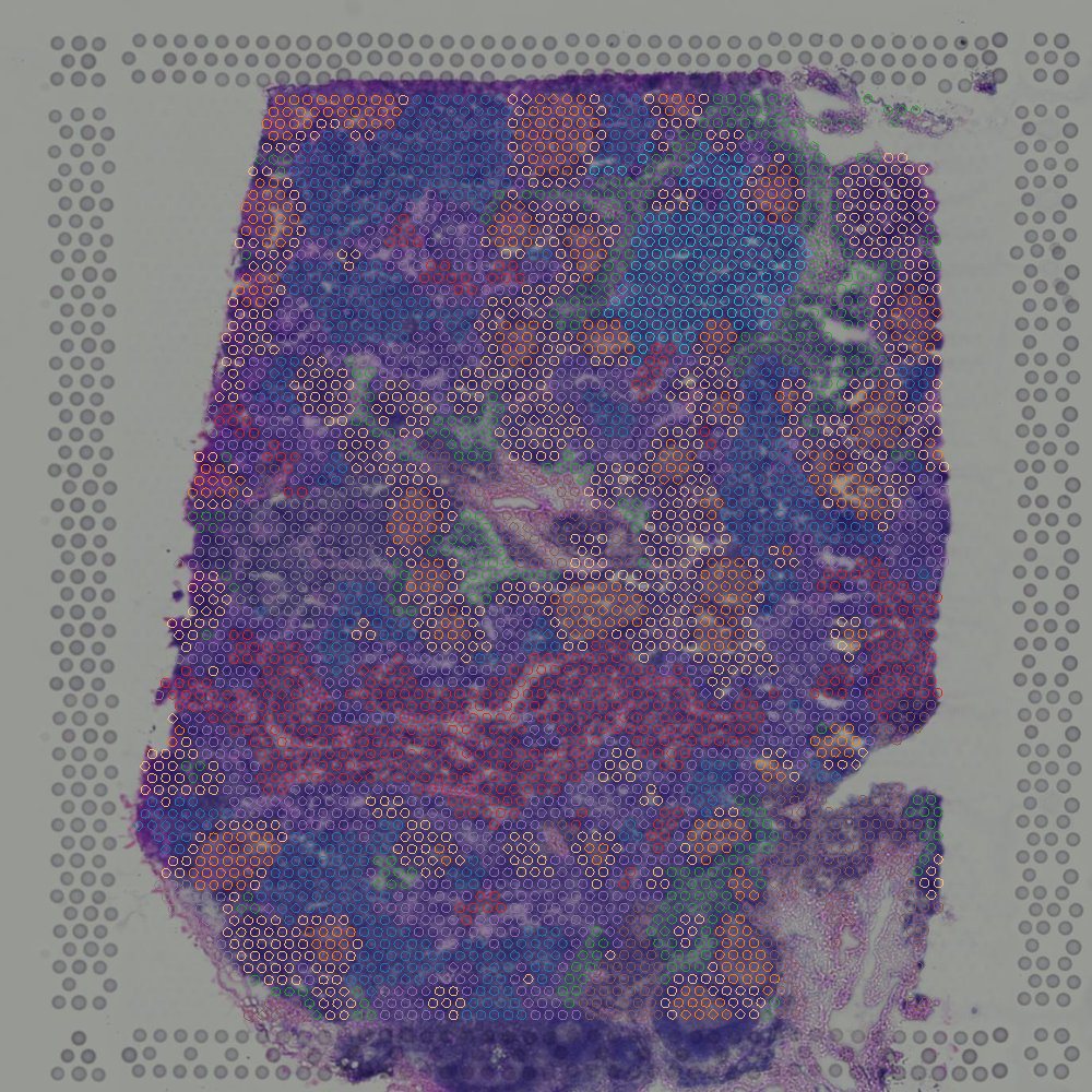
How scientists turn complex spatial multi-omics data into biological insights.
Spatial domain mapping
Human Lymph Node (Space Ranger 1.1.0)
View the analysis in ORAIN (opens in a new tab, no login needed)Spatially variable genes
Human lung cancer (FFPE), immunofluorescence-stained section
View the analysis in ORAIN (opens in a new tab, no login needed)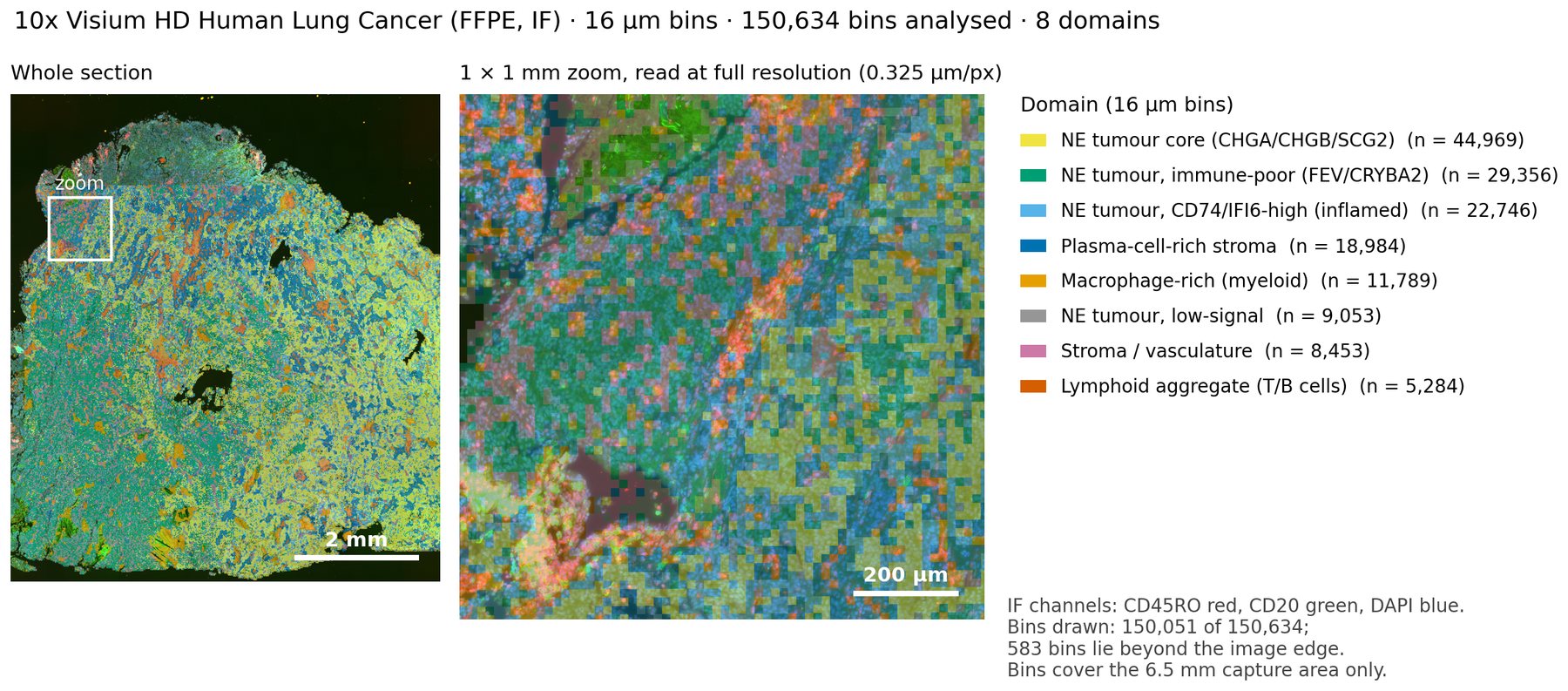
Single-cell spatial typing on H&E
Human lung cancer (FFPE), 289-gene lung panel, with H&E of the same section
View the analysis in ORAIN (opens in a new tab, no login needed)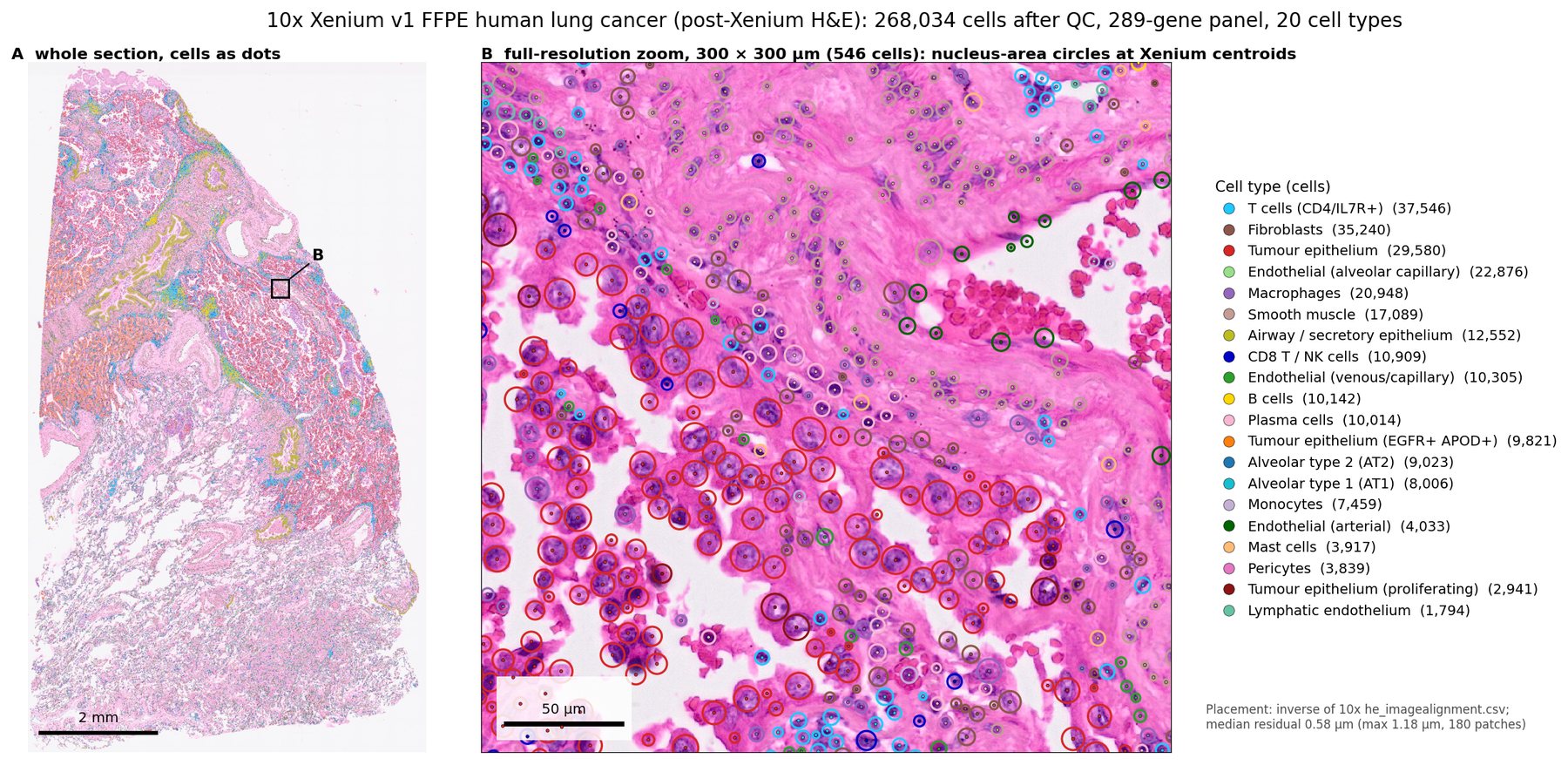
Cell typing across a tissue microarray
Colorectal cancer tissue microarray (MSI and MSS tumors with normal mucosa), 1,000-plex RNA panel
View the analysis in ORAIN (opens in a new tab, no login needed)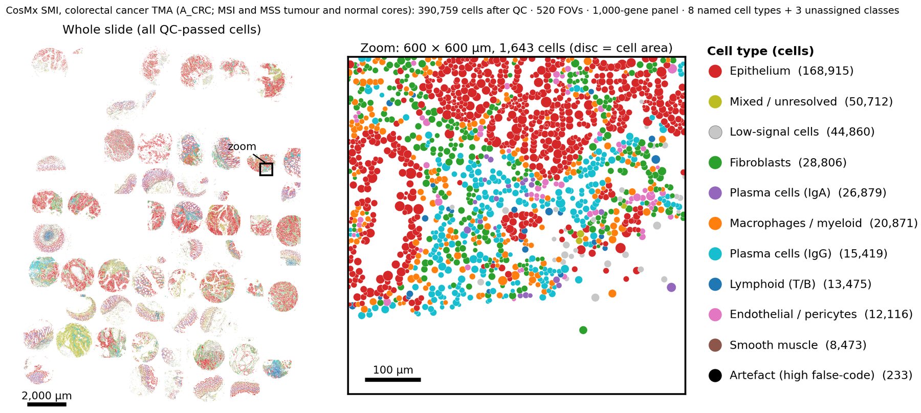
Tumor microenvironment composition
Human ovarian cancer, 316-gene immuno-oncology panel
View the analysis in ORAIN (opens in a new tab, no login needed)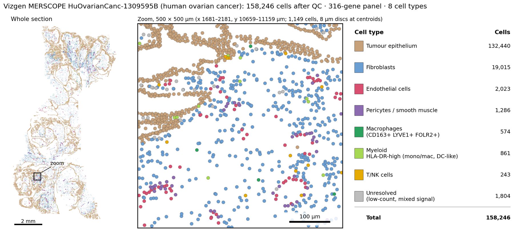
Differential expression between tissue compartments
Human kidney biopsies, minimal change disease vs controls (GSE277674)
View the analysis in ORAIN (opens in a new tab, no login needed)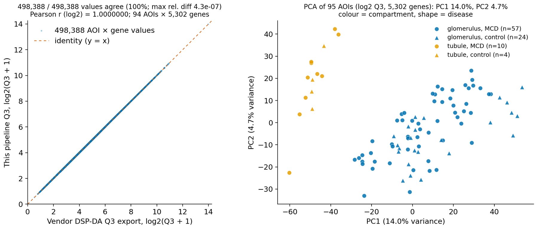
Cell neighborhood analysis
Normal human bone marrow, 54-marker PhenoCycler-Fusion scan (H36)
View the analysis in ORAIN (opens in a new tab, no login needed)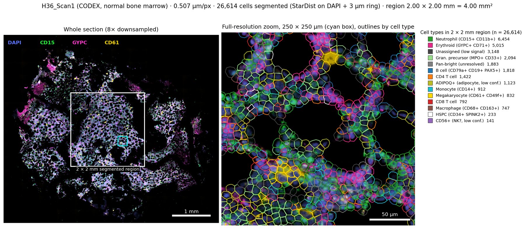
Marker positivity and threshold sensitivity
LuCa-7color whole-slide scan, original Vectra Polaris file
View the analysis in ORAIN (opens in a new tab, no login needed)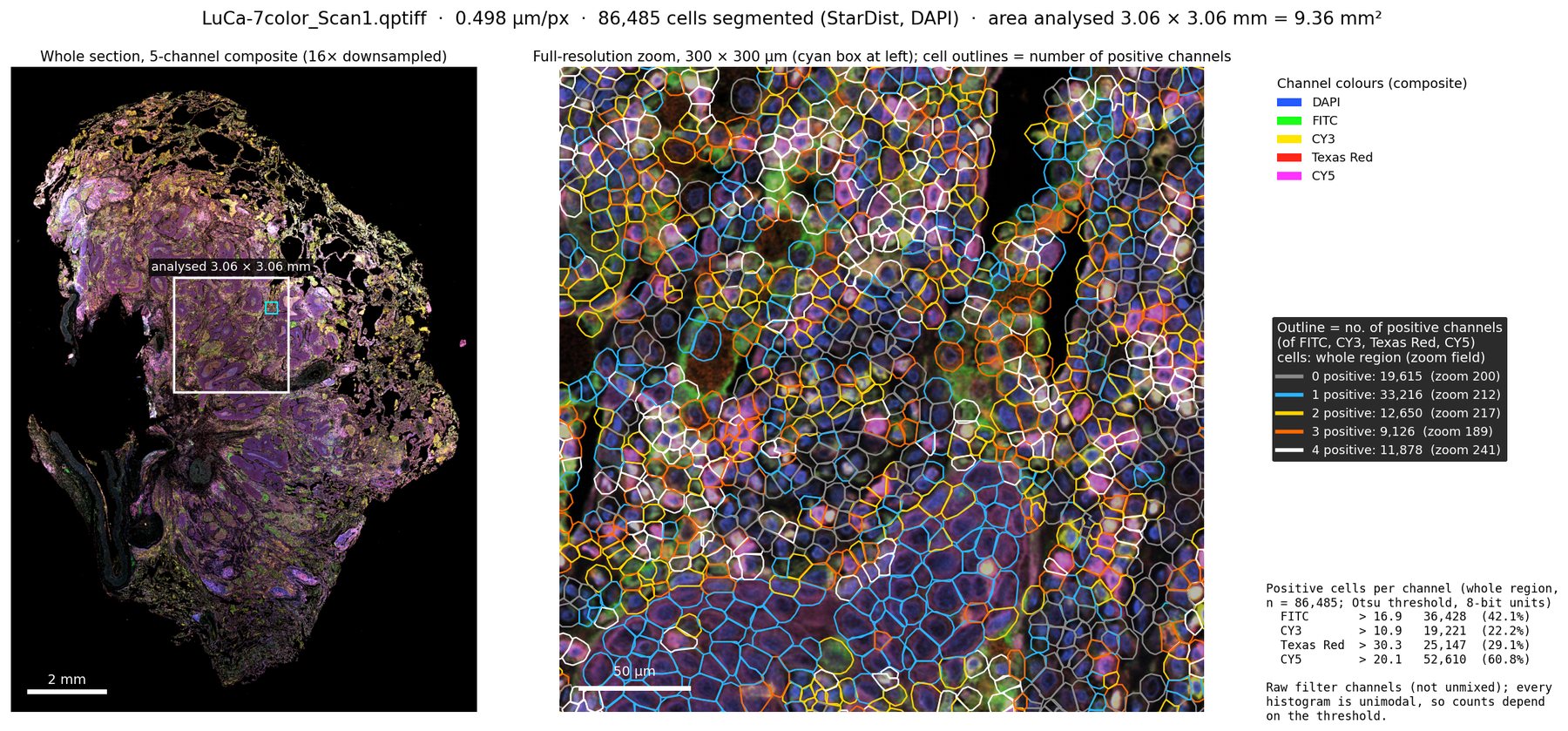
Channel phenotypes and neighborhoods
LuCa-7color whole-slide scan (Vectra Polaris)
View the analysis in ORAIN (opens in a new tab, no login needed)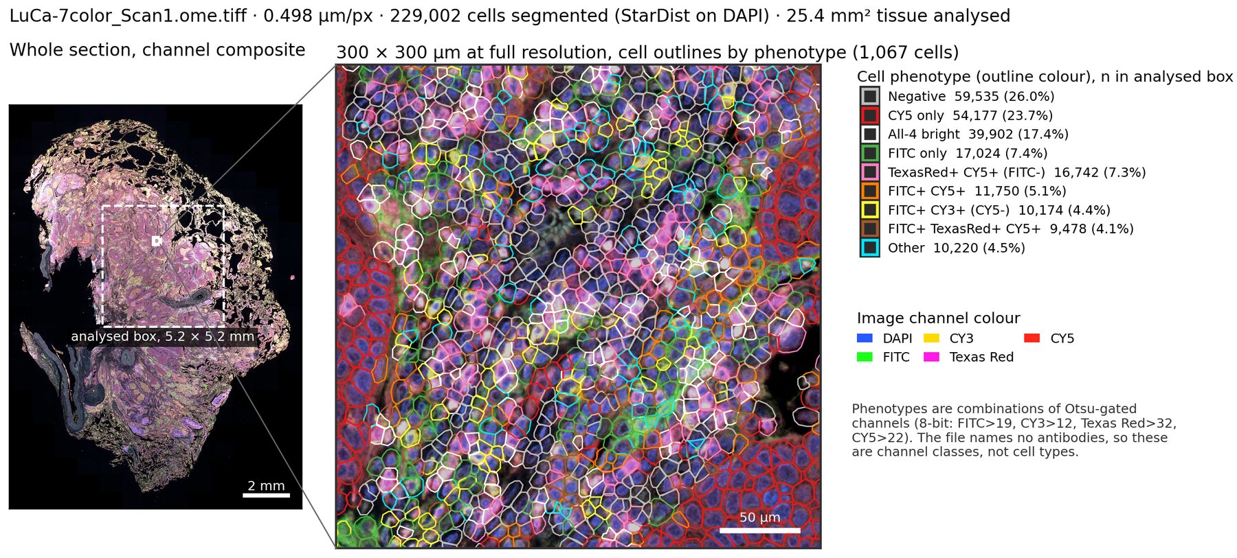
Imaging mass cytometry cell phenotyping
Example imaging mass cytometry raw data, patient 1
View the analysis in ORAIN (opens in a new tab, no login needed)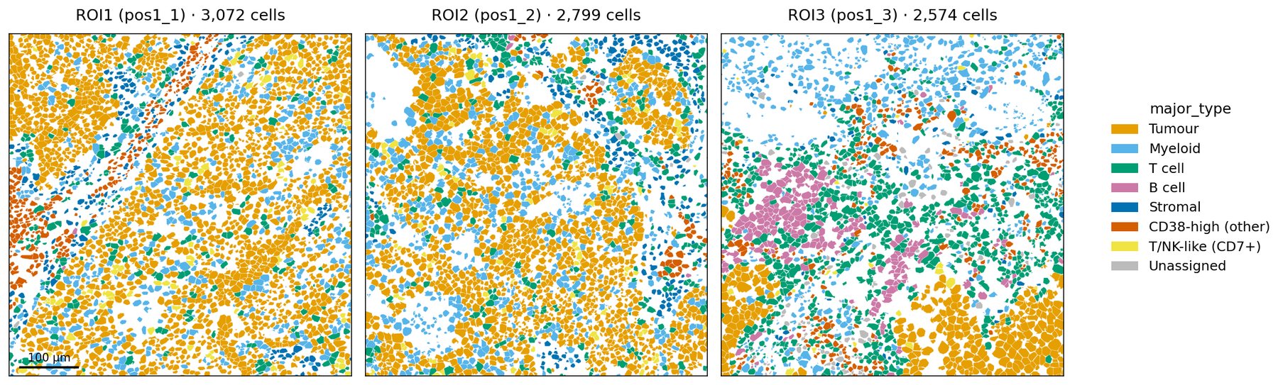
scRNA-seq cell annotation
10k PBMCs from a healthy donor (v3 chemistry)
View the analysis in ORAIN (opens in a new tab, no login needed)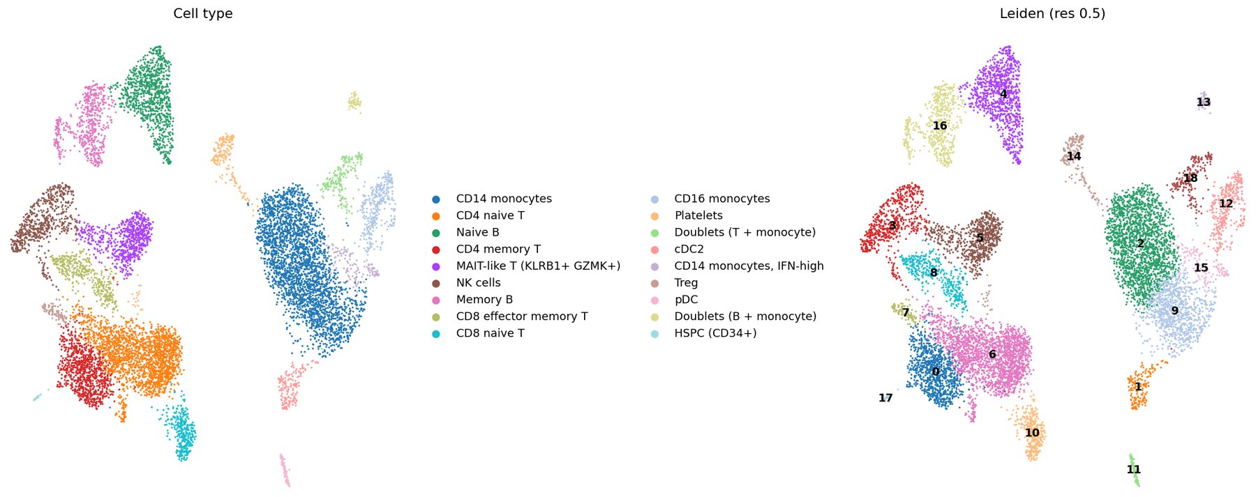
Checking curated cell labels
Immune cells from SCLC patients (Ng et al. 2026, Cancer Cell)
View the analysis in ORAIN (opens in a new tab, no login needed)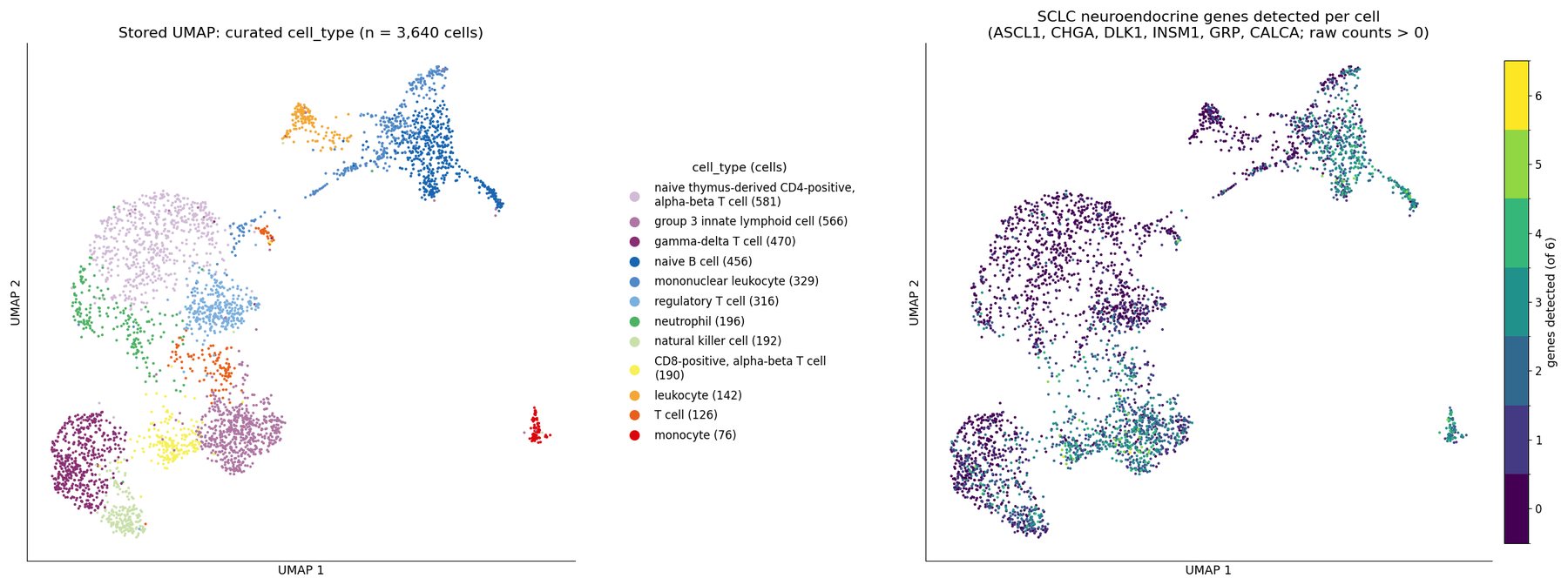
Mutation co-occurrence analysis
TCGA lung adenocarcinoma, PanCancer Atlas: 566 sequenced tumors
View the analysis in ORAIN (opens in a new tab, no login needed)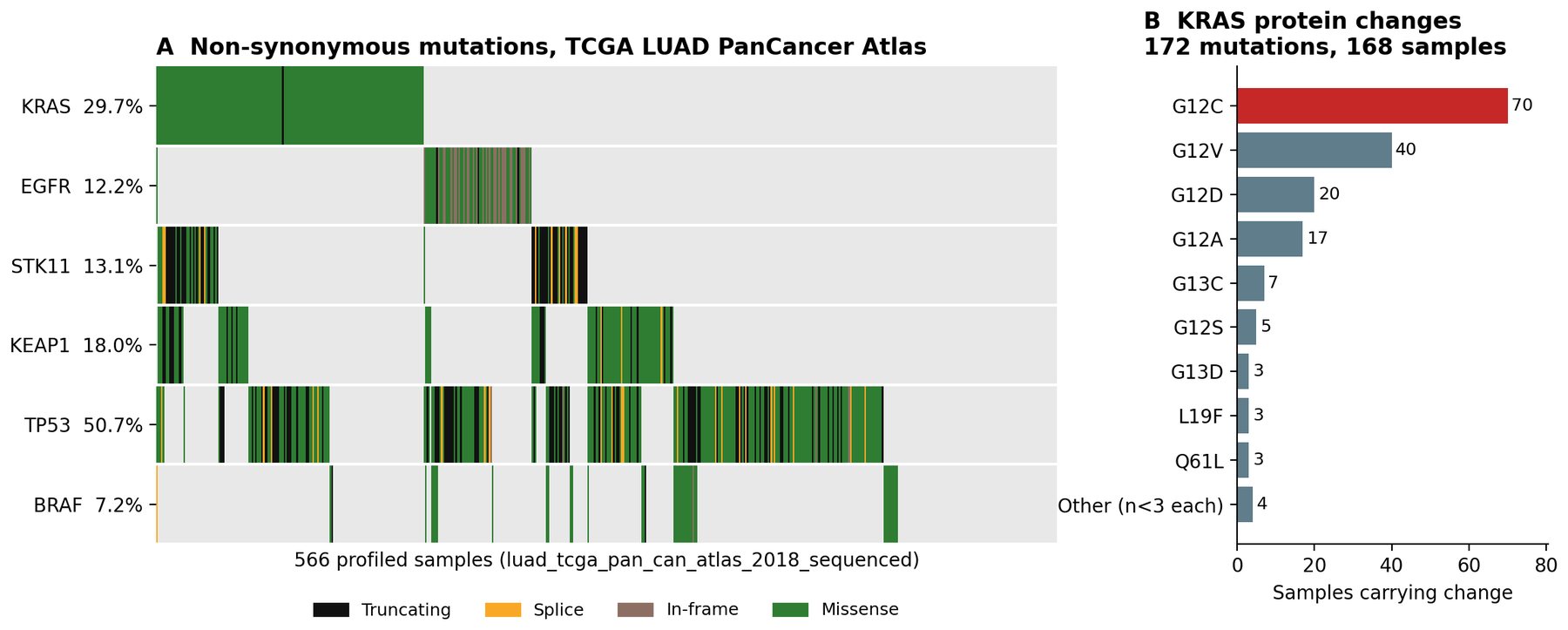
Tissue and nuclei quantification
TCGA-AR-A0TX diagnostic H&E slide (TCGA-BRCA)
View the analysis in ORAIN (opens in a new tab, no login needed)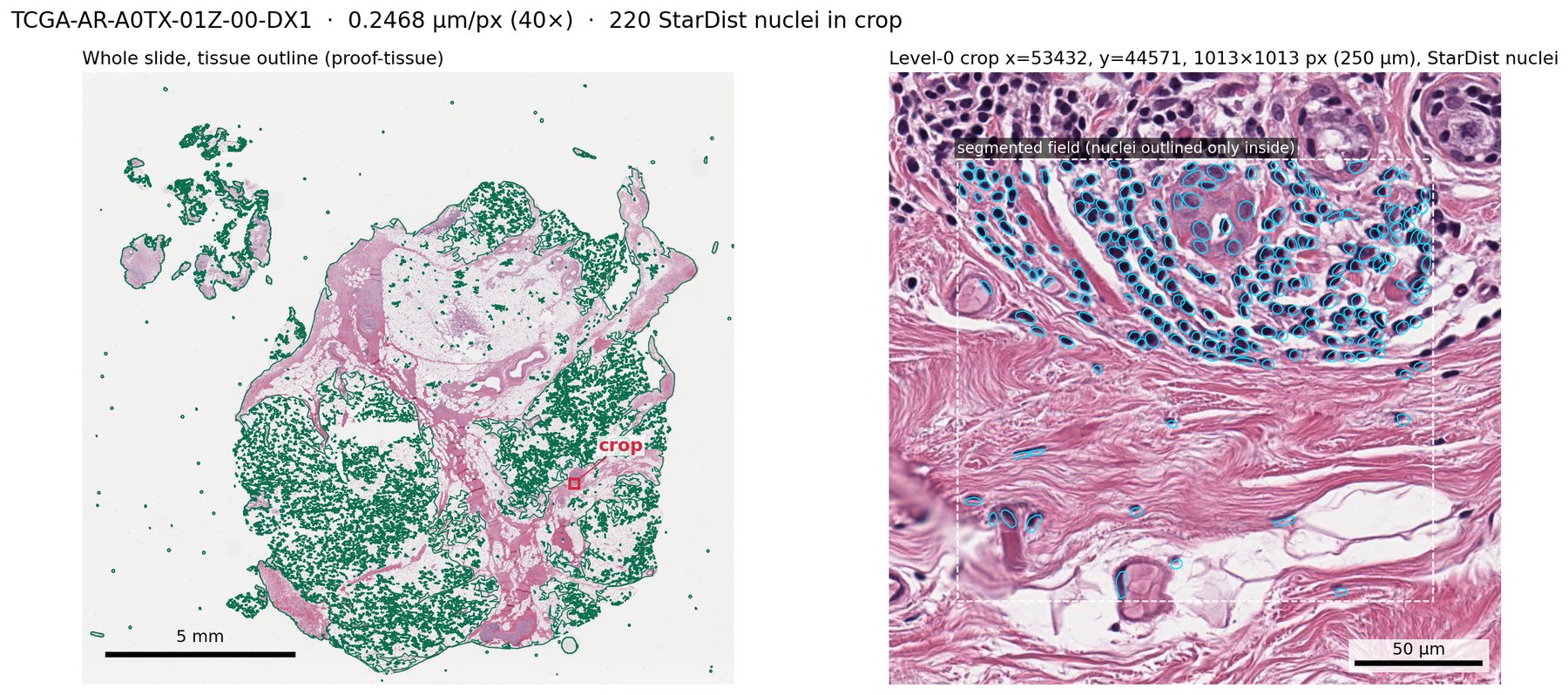
Nuclei density on H&E
Hamamatsu-2: H&E section scanned on a Hamamatsu NanoZoomer
View the analysis in ORAIN (opens in a new tab, no login needed)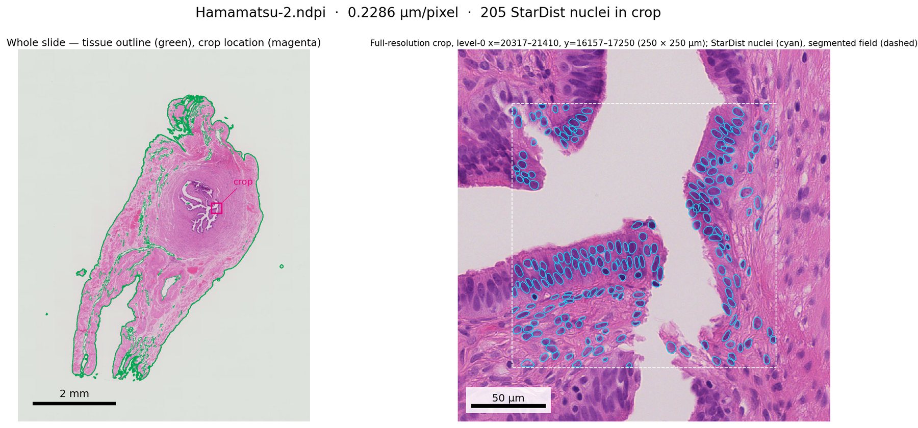
Lymph node cellularity
Philips-1: lymph node H&E from CAMELYON16
View the analysis in ORAIN (opens in a new tab, no login needed)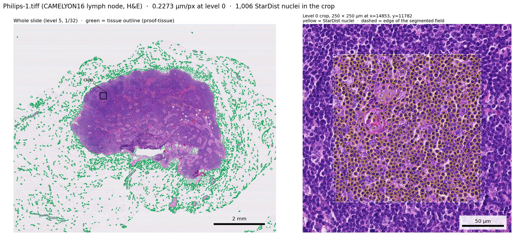
Nuclei counting on trichrome
Zeiss-5: mouse kidney, trichrome stain, two scenes
View the analysis in ORAIN (opens in a new tab, no login needed)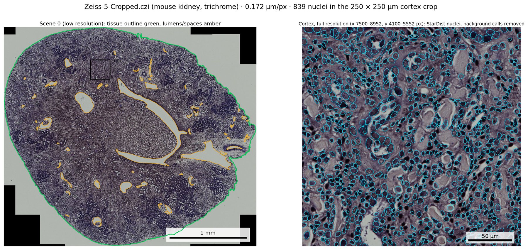
Nuclei density across sections
Leica-2: mouse kidney H&E, four sections scanned as separate 40× regions
View the analysis in ORAIN (opens in a new tab, no login needed)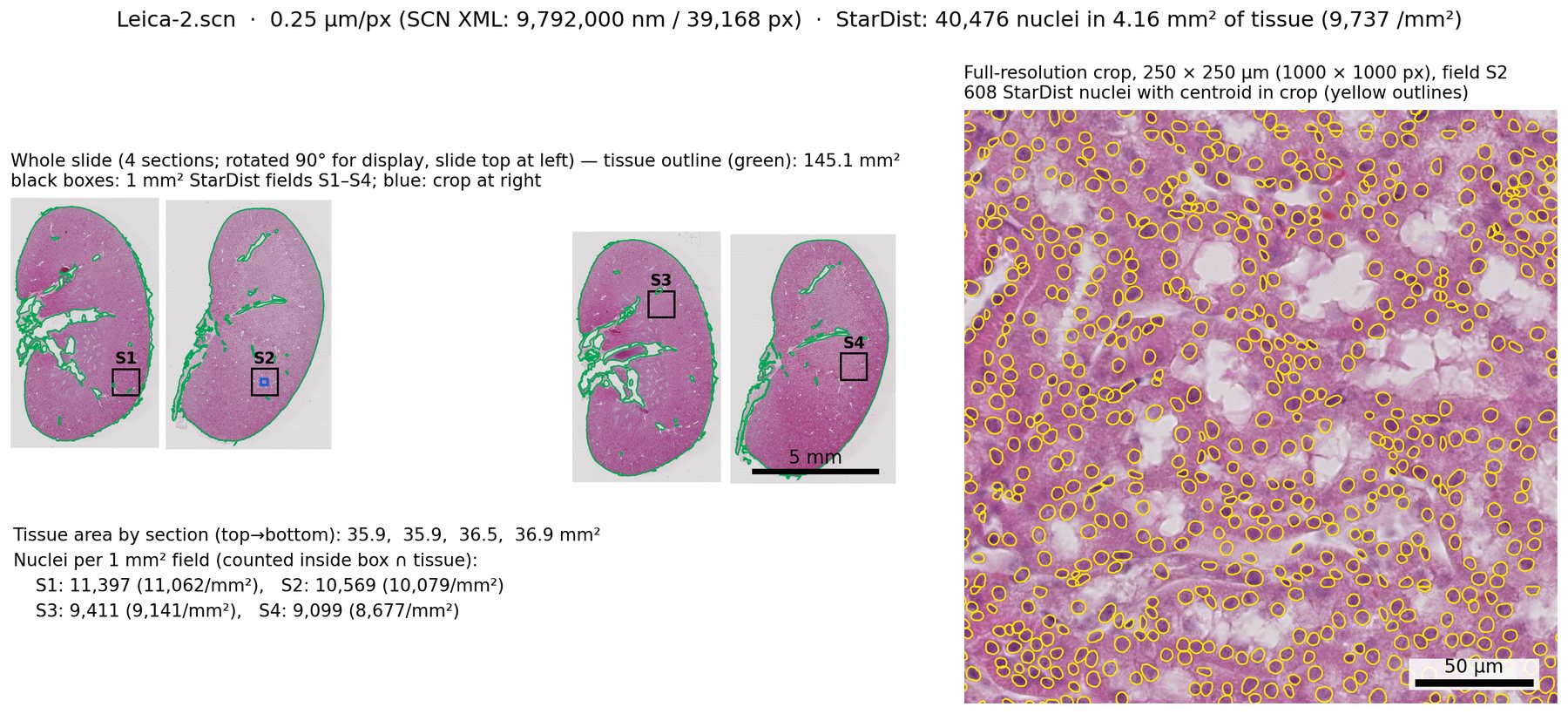
Bring your slides and spatial data to ORAIN, or talk to our scientists about a study.
All inquiries are treated as confidential.